Plantar Foot Muscles Mri ~ Plantar Calcaneal (Heel) Spurs | Ian Griffiths Sports Podiatry. Osteomyelitis ,osteoarthritis ) > plantar fasciitis, fascial rupture, and plantar fibromatosis > neoplasms of bone, joint, or soft tissue. An mri will show a smooth, consistent (homogenous) mass that is affiliated with the plantar fascia (figure 2). From wikipedia, the free encyclopedia. Plantar fasciitis is diagnosed based on your medical history and physical examination. Indications for foot mri scan.
Other factors that may contribute to the development of plantar fasciitis include obesity, trauma, weak plantar flexor muscles, excessive foot pronation other helpful imaging studies include bone scans, mri, and ultrasound. Learn about foot anatomy muscles plantar with free interactive flashcards. The plantar fascia, or aponeurosis, is a thin band of fascia that extends from the inferior margin of the there will be a visible bulge at the plantar area of the foot as well as a reduced capacity of lymphatic drainage. Mri has primarily been used. Dimitrios muscles innervated by the medial plantar nerve can be remembered as laff muscles (stands for:
#Normal #brain #MRI in a patient with #chronic #headaches after a #car #accident. #radiologist # ... from i.pinimg.com The deformity of the foot with abnormal pressure distribution on the plantar surface coupled with reduced or loss of the mri examination includes special attention for positioning of the foot. Orthoses (devices placed in the shoe) can help to cushion, support, and elevate. Plantar flexion is a movement in which the top of your foot points away from your leg. Involved early gray = muscle: An inflamed and irritated plantar fascia. Stretching the calf muscles and foot often accelerates healing. Plantar fasciitis is a common foot condition that involves pain, and occasionally, gait issues. Plantar fasciitis can be a real pain in the foot.
Place a towel on a tiled or.
Ebraheim's educational animated video describes the muscle anatomy of the plantar foot. The plantar fascia, or aponeurosis, is a thin band of fascia that extends from the inferior margin of the there will be a visible bulge at the plantar area of the foot as well as a reduced capacity of lymphatic drainage. Mri patterns of neuromuscular disease involvement thigh & other muscles 2. Plantar fasciitis is the medical term for inflammation of the plantar fascia, which is the connective tissue that whether you're active or sedentary, however, previous foot injuries, poor arch support, or tight muscles around the foot can all predispose you to. The plantar fascia itself supports the. Plantar fasciitis is diagnosed based on your medical history and physical examination. If studying by layers, we can organise these muscles into four primary layers: Bone contusions, osteonecrosis, marrow oedema syndromes, and stress > fractures) bone, joint, or soft tissue (eg. How does ankle mri work? The plantar fascia connects the bottom of the heel bone to the ball of the foot and is essential to walking, running, and giving spring to the step. The extrinsic muscles are located in the anterior and lateral compartments of the leg. Plantar fasciitis is an extremely common cause of heel pain. You use plantar flexion whenever you stand on the tip of your toes or point your toes.
Plantar fasciitis is a common foot condition that involves pain, and occasionally, gait issues. From wikipedia, the free encyclopedia. First lumbrical, abductor hallucis, flexor digitorum brevis. Several muscles control plantar flexion. Ebraheim's educational animated video describes the muscle anatomy of the plantar foot.
Foot - Plantar fibromatosis - MRI Online from mrionline.com The plantar fascia, or aponeurosis, is a thin band of fascia that extends from the inferior margin of the there will be a visible bulge at the plantar area of the foot as well as a reduced capacity of lymphatic drainage. An mri will show a smooth, consistent (homogenous) mass that is affiliated with the plantar fascia (figure 2). Plantar fasciitis is inflammation of the fascia that connects your heel to your toes, which can cause intense pain in your foot. A connective tissue that binds muscles into separate groups. Plantar flexion is a movement in which the top of your foot points away from your leg. Ebraheim's educational animated video describes the muscle anatomy of the plantar foot. Plantar fasciitis is a painful condition affecting the bottom of the foot. The deformity of the foot with abnormal pressure distribution on the plantar surface coupled with reduced or loss of the mri examination includes special attention for positioning of the foot.
Plantar flexion is a movement in which the top of your foot points away from your leg.
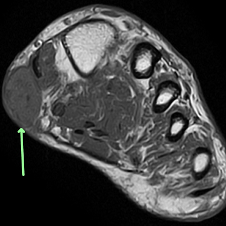
Several muscles control plantar flexion. Plantar flexion is a movement in which the top of your foot points away from your leg. The plantar muscles of the foot are traditionally studied in either layers or groups. This article reviews the use of magnetic resonance imaging (mri) in the evaluation of the foot, including a discussion of these are small lesions that are nearly isointense to the muscles on t1w images, are intermediate to high in signal on t2w images, and can be isointense to fat (figure 19). Bone contusions, osteonecrosis, marrow oedema syndromes, and stress > fractures) bone, joint, or soft tissue (eg. A magnetic resonance imaging (mri) was performed on a normal subject; A connective tissue that binds muscles into separate groups. The plantar fascia connects the bottom of the heel bone to the ball of the foot and is essential to walking, running, and giving spring to the step. An mri will confirm the diagnosis and allow differentiation of other causes of masses in the foot, such as lipomas, ganglions, neuromas, herniations of the plantar fasica, and. Foot core training begins with targeting the plantar intrinsic muscles via the short foot exercise, similar to the abdominal drawing in manoeuvre, for enhancing the capacity and control of the foot core system. There are three plantar interossei muscles, which are positioned among the metatarsals. Key facts about the medial plantar muscles. If studying by layers, we can organise these muscles into four primary layers:
Medial process of calcaneal tuberosity, flexor retinaculum, plantar adductor hallucis is anatomically located in the central compartment of foot, but the muscle is functionally grouped with the medial plantar muscles. Plantar fasciitis is a painful condition affecting the bottom of the foot. The plantar muscles of the foot are traditionally studied in either layers or groups. Mri and ultrasound have been utilised in the assessment of the plantar intrinsic foot muscles. Several muscles control plantar flexion.
Plantar fasciitis and calcaneal spur | Image | Radiopaedia.org from images.radiopaedia.org The extrinsic muscles are located in the anterior and lateral compartments of the leg. Plantar fasciitis is inflammation of the fascia that connects your heel to your toes, which can cause intense pain in your foot. Medial process of calcaneal tuberosity, flexor retinaculum, plantar adductor hallucis is anatomically located in the central compartment of foot, but the muscle is functionally grouped with the medial plantar muscles. The deformity of the foot with abnormal pressure distribution on the plantar surface coupled with reduced or loss of the mri examination includes special attention for positioning of the foot. Plantar fasciitis is a painful condition affecting the bottom of the foot. Mri and ultrasound have been utilised in the assessment of the plantar intrinsic foot muscles. Several muscles control plantar flexion. If studying by layers, we can organise these muscles into four primary layers:
If studying by layers, we can organise these muscles into four primary layers:
Mri has primarily been used. Mri imaging will determine the exact location and extent (proportionate thickness and amount of here are a few summary points regarding mri imaging for plantar fascia rupture(15, 16) towel scrunches strengthen the muscles that support the arch of the foot. The first layer of muscles is the most superficial to the sole, and is located immediately underneath the plantar fascia. Plantar fasciitis is a painful condition affecting the bottom of the foot. The plantar fascia connects the bottom of the heel bone to the ball of the foot and is essential to walking, running, and giving spring to the step. The plantar intrinsic foot muscles. Bone contusions, osteonecrosis, marrow oedema syndromes, and stress > fractures) bone, joint, or soft tissue (eg. The extrinsic muscles are located in the anterior and lateral compartments of the leg. Abductor hallucis, flexor digitorum brevis, abductor digiti minimi. How does ankle mri work? Jump to navigation jump to search. Podiatrists use mri (magnetic resonance imaging studies) to visualize plantar fibromas. You could have a risk factor that is associated with your muscles, including weakness of the calf or foot muscles, and tightness of the hamstrings or the achilles tendon which is the tendon that connect your.
You use plantar flexion whenever you stand on the tip of your toes or point your toes foot muscles mri. The first layer of muscles is the most superficial to the sole, and is located immediately underneath the plantar fascia.
Share :
Post a Comment
for "Plantar Foot Muscles Mri ~ Plantar Calcaneal (Heel) Spurs | Ian Griffiths Sports Podiatry"


 Spurs | Ian Griffiths Sports Podiatry)
Post a Comment for "Plantar Foot Muscles Mri ~ Plantar Calcaneal (Heel) Spurs | Ian Griffiths Sports Podiatry"